As creatures get more complex, the light in their myelin shifts red
In a study published in 2009, to photograph a man by his own light, Masaki Kobayashi and his colleagues in Sendai first took away all other light. The volunteer sat in the dark room for fifteen minutes before anything was recorded. Then a camera cooled far below freezing looked at him for twenty minutes.
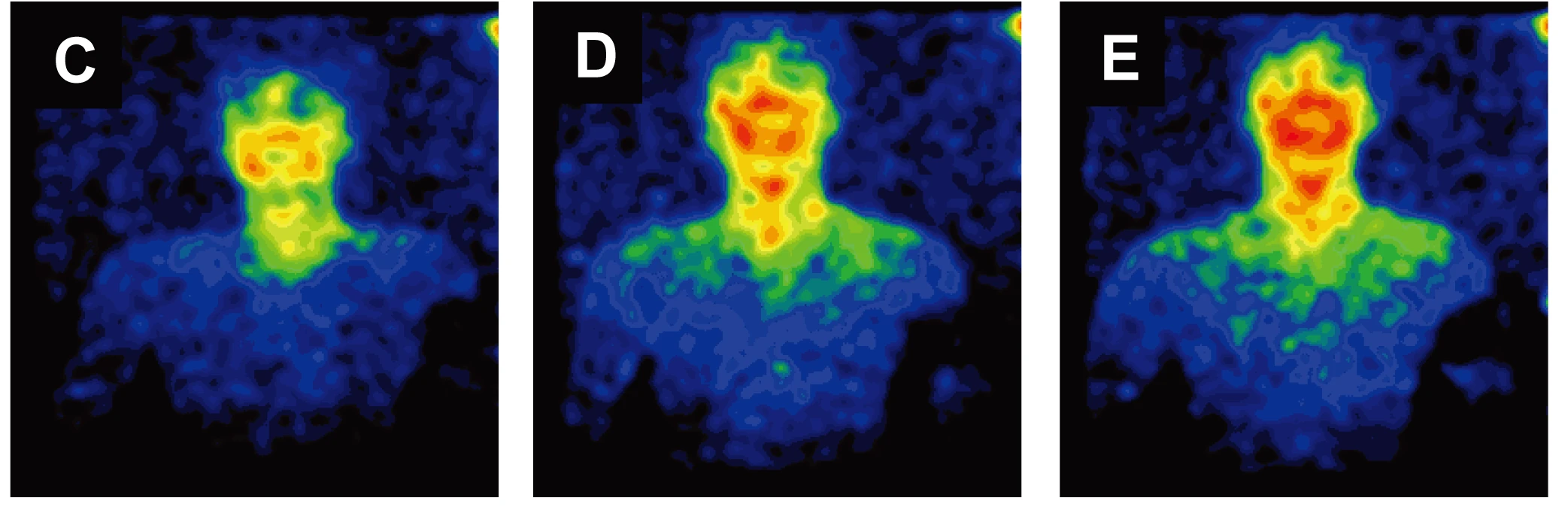
It collected a few hundred photons per square centimetre per second, about a thousand times less than the eye can see. The face was brighter than the body, the mouth and cheeks brighter than the rest of the face, and the whole figure was brightest around four in the afternoon.
These photons are called biophotons, or, more soberly, ultraweak photon emission. There is nothing occult in them. They are a by-product of chemistry. Oxidation in the cell leaves some molecules in an excited state, and a few of them return to rest by giving up a photon. All living tissue does it. The brain does it too, and the brain is full of a structure that an optician would recognise.
The sheath
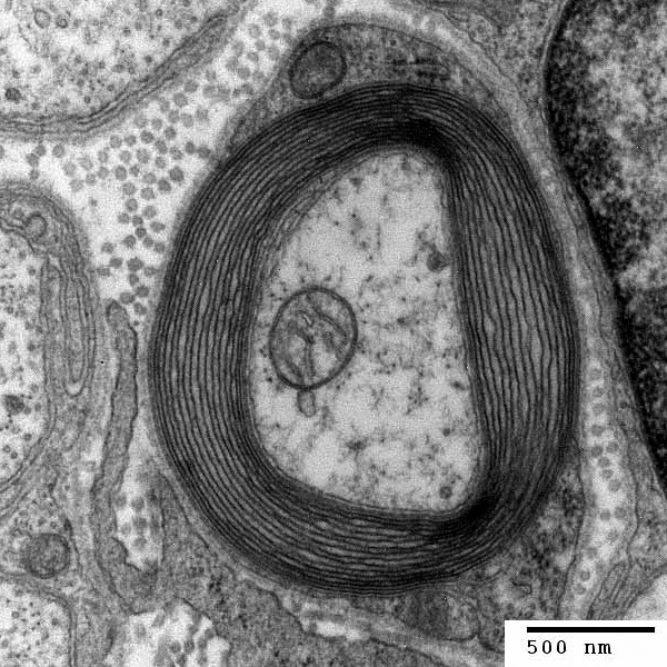
A myelinated axon is a thin thread of cytoplasm around which another cell has wound its own flattened membrane, many times, like paper rolled tight around a pencil. The wraps are mostly fat.
Their refractive index is higher than that of the axon inside and of the fluid outside. Light travelling in a denser layer, at a shallow enough angle, is reflected at the boundary and stays in. An optical fibre works the same way.
In 2016 Sourabh Kumar, Christoph Simon and their colleagues calculated that a myelin sheath could carry biophotons this way along an axon, even when the axon bends and the sheath is uneven.

Six brains
The same year, in Wuhan, Zhuo Wang, Jiapei Dai and their colleagues measured the colour of the biophotons that brains give off. They kept slices of brain alive in solution and gave them glutamate, the transmitter that excites most neurons. The faint light passed through a slit and a grating onto a camera cooled to −95 °C, and its spread on the sensor gave its wavelengths.
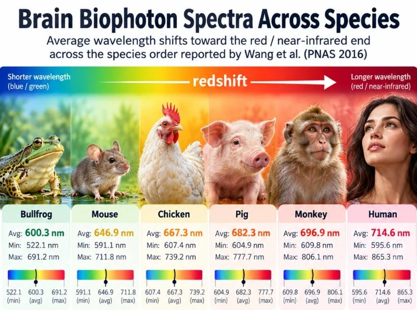
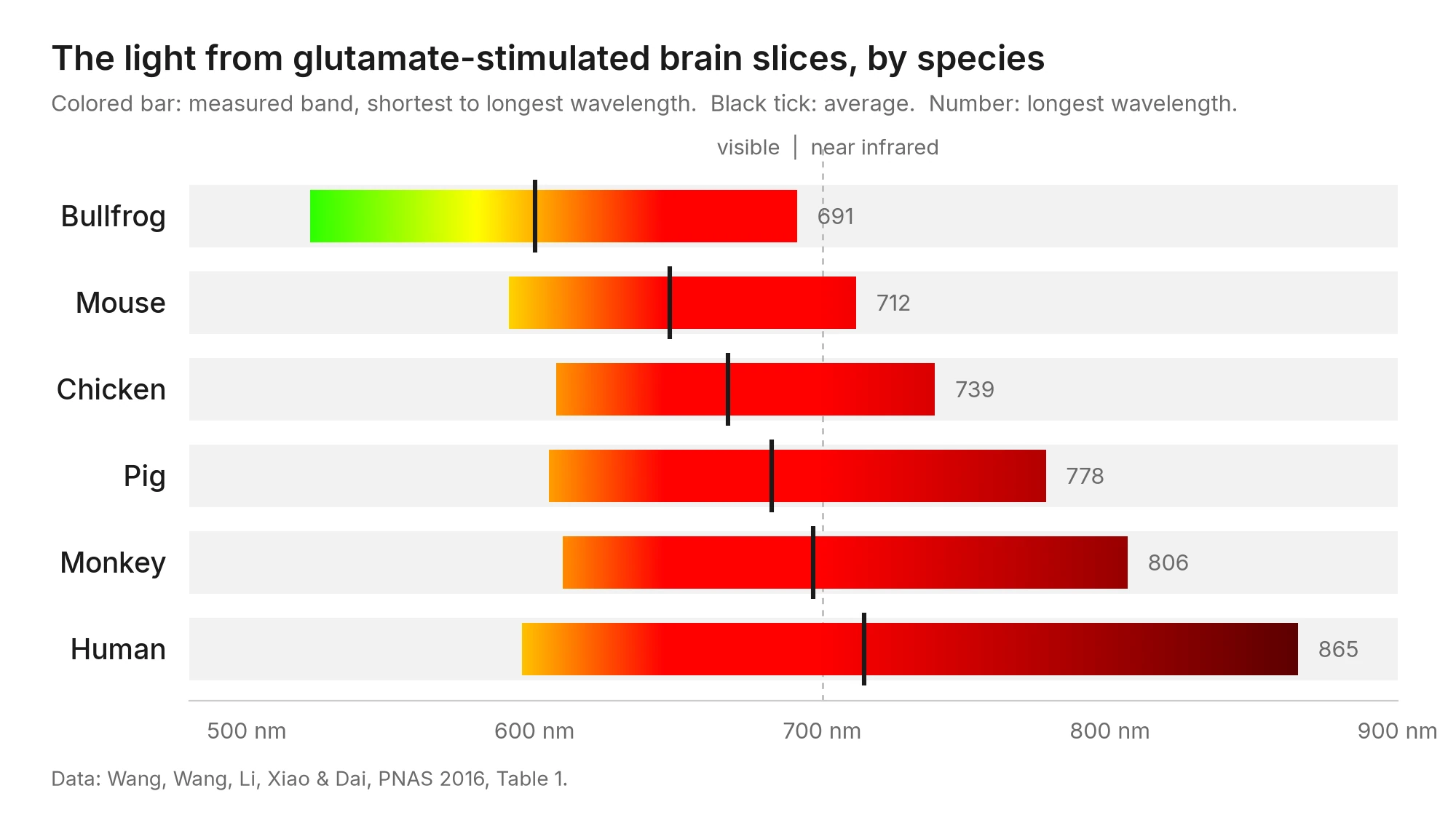
They did this for six animals. In the bullfrog the light ran from 522 to 691 nanometres, from green through orange to red. In human tissue it ran from 596 to 865, well past the red into the infrared, where the eye sees nothing. Mouse, chicken, pig and monkey fell between them, in that order.
Drawn to scale, the bands show something the averages hide. The short end hardly moved. The long end moved out.
The number of turns
A colleague put the idea to me in one line:
There could be a relationship between nervous system architecture and the spectral properties of biophotons.
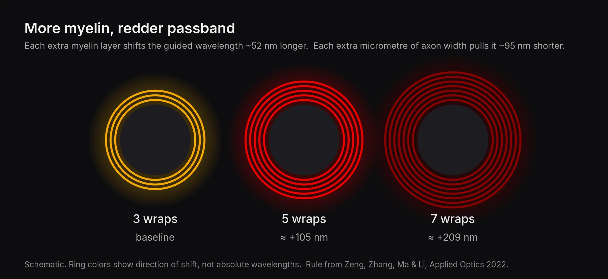
One such relationship has been calculated. In 2022 Haomin Zeng and colleagues, also in Wuhan, worked out how light would travel in such a sheath. They found it carries only a narrow band of colours, about ten nanometres wide, and that the band is set by the shape:
One more layer of myelin moves the band about 52 nanometres toward the red.
An axon one micrometre wider moves it about 95 nanometres toward the blue.
The colour depends on the number of turns, as the note of a string depends on its length. They proposed this as the reason for the difference between the six brains.
Age
With age the shift goes the other way. In mice from newborn to eighteen months, the same group found the light moving back toward the blue. In old mice myelin breaks down and is renewed more slowly.
A red photon carries less energy than a blue one, and the authors read the change as an ageing brain spending more on each signal.
It is a strange thought, that age might show as a change of colour in a light no one can see.
Intelligence
The Wuhan group go further and present the redshift as a physical basis for human intelligence. Here the evidence thins. A reply in the same journal pointed out that the wavelengths also follow body mass, and that the human light had the shortest coherence length of the six. The slices were bathed in 50 millimolar glutamate, far more than any living brain receives. And the chicken came out redder than the mouse, which may say more about birds than about the theory.
The receiver
A signal needs something to read it, and no experiment has yet shown a neuron taking in such a photon and acting on it. But the brain is not without eyes of its own.
In 1999 a light-sensitive protein, encephalopsin, was found spread through the mammalian brain: in the hypothalamus, in parts of the cortex, in the Purkinje cells of the cerebellum, places that never see the sky. Birds go further. In the quail, neurons deep in the hypothalamus carry an opsin called OPN5. They register the daylight that passes through feathers, skin and bone, and the bird uses it to tell the season.
The receivers described so far answer to violet and blue, not to the red and infrared that brain tissue gives off. If something reads the light in the sheaths, it has not been found. But the parts are all there: a source in every cell, a fibre around every fast nerve, and proteins in the dark that respond to light.
Each of us gives off a little light, most of it from the face, most of it in the afternoon. Inside the skull, some of those biophotons may run along sheaths of fat wound around the nerves, their colour fixed by how many times they are wound.
Whether anything in that darkness reads them, we do not know.